


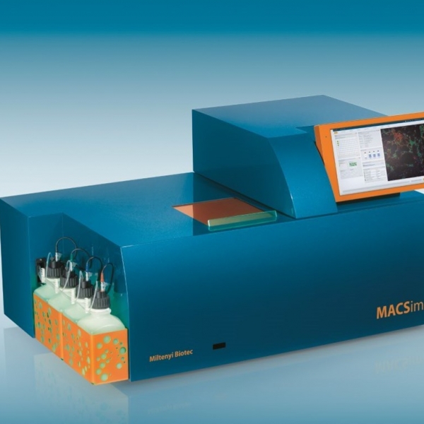
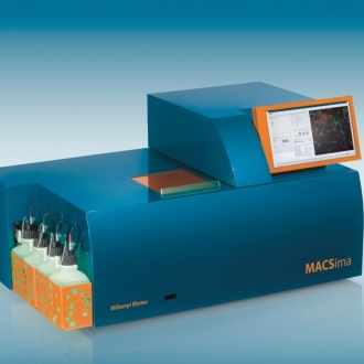
MACSima™ Imaging Platform
MACSima™ Imaging Platform - The easy way to ultrahigh-content imaging
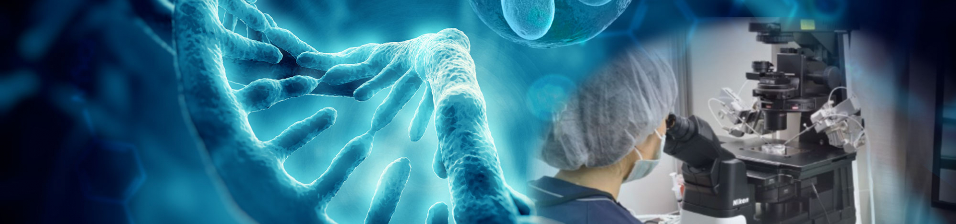
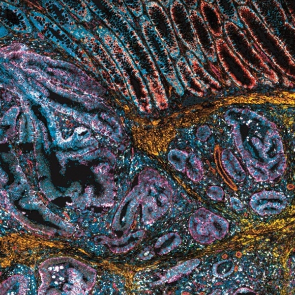
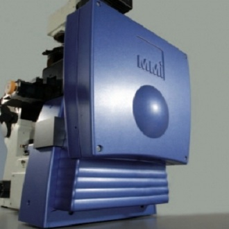
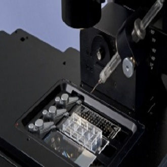
The MACSima Imaging Platform can enables fully automated high-content imaging. The possibility of evaluating localization, expression, and potential interaction of a multitude of different proteins allows scientists to tap the full potential of spatial biology.
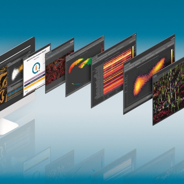
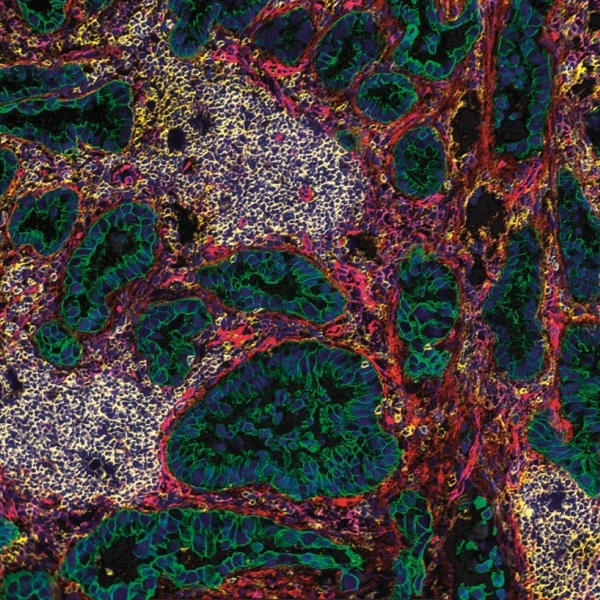
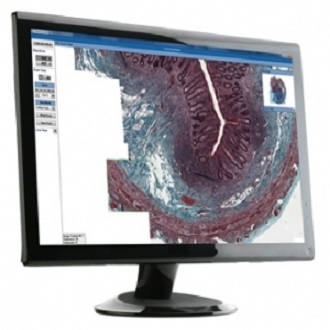
MACSima Imaging Cyclic Staining (MICS) technology enables the simultaneous analysis of hundreds of markers on a single sample based on fluorescence microscopy. It uses the principle of iterative staining with different fluorochrome-conjugated antibodies to acquire microscopy data for a multitude of parameters without harming the sample. The iterative process comprises three main steps: fluorescent staining, image acquisition, and erasure of the fluorescence signal, all of which are conducted by the MACSima Imaging System in a fully automated manner. The resulting stack of potentially hundreds of marker images provides an unprecedented insight into the physiological or pathological characteristics of the sample. Due to on-the-fly processing, data analysis can start at any time, even when the iterative process is still running.
The MACSima Imaging Platform can enables fully automated high-content imaging. The possibility of evaluating localization, expression, and potential interaction of a multitude of different proteins allows scientists to tap the full potential of spatial biology.
MACSima Imaging Cyclic Staining (MICS) technology enables the simultaneous analysis of hundreds of markers on a single sample based on fluorescence microscopy. It uses the principle of iterative staining with different fluorochrome-conjugated antibodies to acquire microscopy data for a multitude of parameters without harming the sample. The iterative process comprises three main steps: fluorescent staining, image acquisition, and erasure of the fluorescence signal, all of which are conducted by the MACSima Imaging System in a fully automated manner. The resulting stack of potentially hundreds of marker images provides an unprecedented insight into the physiological or pathological characteristics of the sample. Due to on-the-fly processing, data analysis can start at any time, even when the iterative process is still running.
Related Products
-
 New
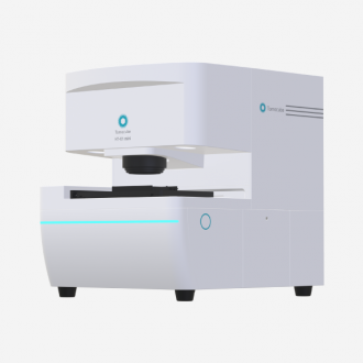
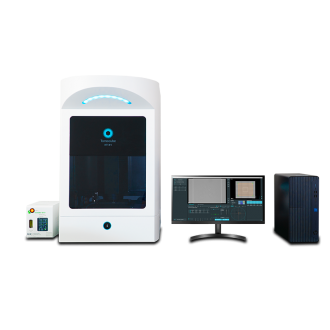
NewHT-X1™ mini : Holotomography I…
-
 New
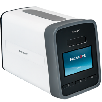
NewFACSCOPE® B
-
 New
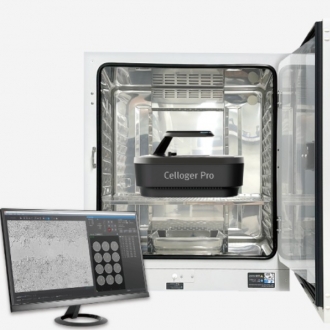
NewCelloger®
-
 New
NewMACSima™ Imaging Platform
-
 Hot
HotTomocube HT-X1
-
 New
NewDigitail
-
 New
NewLabstamp
-
 New
NewIntravital Microscopy
-
 Hot
HotCA Series - Multiple Output Em…
-
Confocal and Multiphoton micro…
-
 New
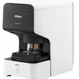
NewNikon Eclipse Ui
-
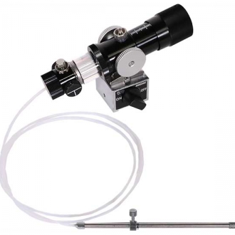
IM-21 Microinjector
-
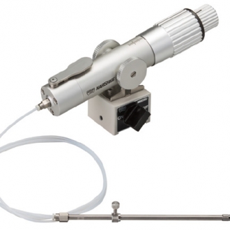
IM-11-2 Pneumatic Microinjecto…
-
ONGO SPERM
-
Injection, holding and biopsy …
-
INTEGRAL HSV
-
DIVA
-
SYMS III sealer
-
Digitcool freezers
-
 Hot
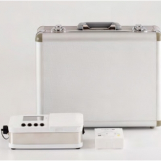
HotMN-2 Mini Bench Top Incubator
-

EMBRYO SHIELD – INLINE FILTER
-
MV60 Wireless Multi-viewing, W…
-
Biopsy micropipettes
-
ICSI Micropipettes
-
Holding Micropipettes
-
DENU-Tips
-
 New
NewDigital Sight Cameras for Micr…
-
Nikon ECLIPSE Ci-L Plus, Uprig…
-
 Hot
HotiXon Ultra 897 facilitates a n…
-
 Hot
HotZL41 Cell sCMOS camera offer a…
-
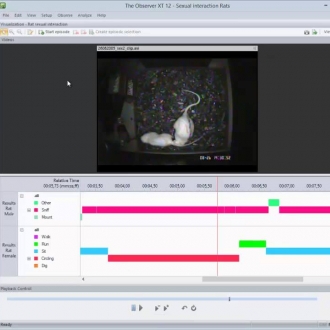
The Observer XT : Professional…
-
by PET, SPECT, Optical Imaging…
-
 New
NewUltraMicroscope Blaze: The aut…
-
 Hot
HotDigital Classroom
-
EthoVision XT: Animal Behavior…
-
Digital Sight Cameras for Micr…
-
 New
NewDigital Sight Cameras for Micr…
-

Shedding New Light On Microsco…
-
Nikon ECLIPSE LV100ND POL/DS P…
-
Nikon ECLIPSE LV100N POL Polar…
-
Smart Station SS-250
-
Nikon ECLIPSE Ci POL Polarized…
-
Nikon ECLIPSE E200 POL Educati…
-
Nikon ECLIPSE Ti2 Inverted Res…
-
Nikon ECLIPSE Ts2R Inverted Re…
-
Nikon ECLIPSE Ts2 Inverted Rou…
-
Nikon ECLIPSE Ni Upright Micro…
-
Nikon ECLIPSE Ci Upright Micro…
-
 New
NewNikon ECLIPSE Si Upright Micro…
-
TissueScope™ LE
-

IVOS II : Computer Asissted Sp…
-
 New
NewNikon Eclipse Ei educational m…
-
sidtest1
-
Water Jacket Penguin AQ AP ser…
-
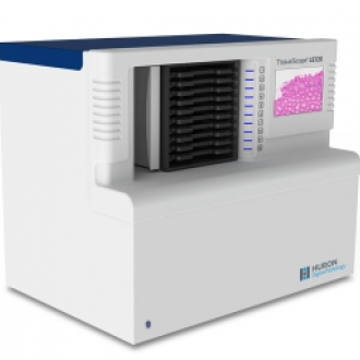
TissueScope™ LE120
-
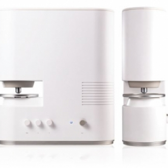
Holotomography Microscope
-
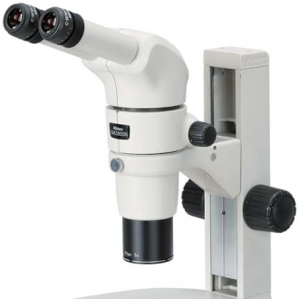
Stereo Microscope (Paralell) S…
-
Panoptiq 3
-
Cell Cut
-
Cellector
-
Xvivo 3